Getting Diagnosed
Talk to your GP if you think your symptoms could be caused by Microscopic Colitis. Your GP will need to take a history of all your symptoms. This may help them rule out other conditions.
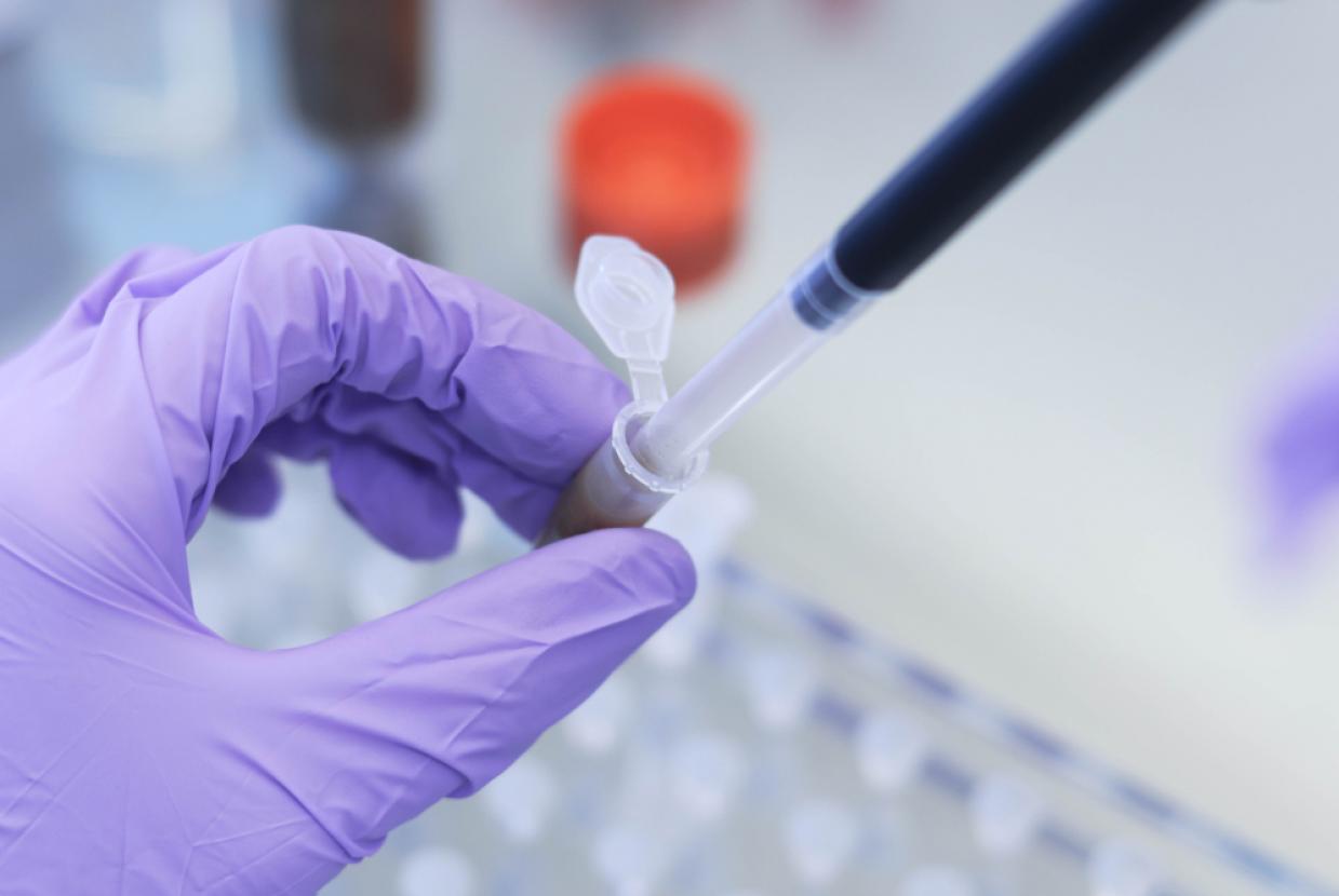
Poo tests, known as faecal calprotectin tests, are often used to help diagnose Crohn’s and Colitis. This test may not be useful for Microscopic Colitis. In Microscopic Colitis, calprotectin levels are often normal or only slightly raised.
If your GP thinks you may have Microscopic Colitis, you will need to be referred for a colonoscopy and a biopsy. You may also need to be tested for other autoimmune conditions, such as coeliac disease.
It may take time to diagnose Microscopic Colitis. This is because the condition is not always as well-recognised as other forms of Inflammatory Bowel Disease.
See our appointment guide for information on getting the most out of your appointments. This includes tips about talking to your healthcare professional.
Colonoscopy with biopsy
In Microscopic Colitis, signs of inflammation can only be seen under a microscope. You will need to have a colonoscopy so that a tissue sample, or biopsy, can be taken.
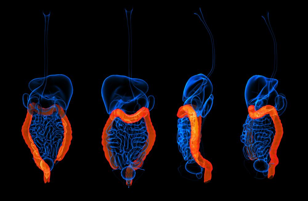
During a colonoscopy, a long flexible tube is inserted through your bottom. The tube is about the thickness of your little finger, with a bright light and camera at its tip, This lets the doctor look at the lining of your colon. During the procedure, a biopsy is taken from your colon. This is then looked at under a microscope.
Most colonoscopies are performed under mild sedation. This is when you are given medicine through a tube in your arm to make you feel relaxed. You may be offered gas and air or painkillers instead, to make you more comfortable.
Other tests you may have
SeHCAT scan
You will be given a small capsule of synthetic bile salts to swallow. This contains a small amount of harmless radioactive material known as SeHCAT. You will then have a scan, followed by another one a week later. These will measure how the bile salts have been absorbed.
See our information on tests and investigations for more on SeHCAT scans. And there is more on bile acid malabsorption in our information on diarrhoea.
Delay in getting a diagnosis
It may take time to get a diagnosis of Microscopic Colitis, which can be frustrating.
This may be because:
- A poo test, or faecal calprotectin test is not always helpful in finding Microscopic Colitis
- Your large bowel looks normal at colonoscopy. A biopsy is needed to find Microscopic Colitis. If your GP or specialist thinks you may have Microscopic Colitis, they will arrange for you to have a biopsy.
- Your symptoms may be similar to conditions such as IBS or coeliac disease. You may need to have a blood test to rule out coeliac disease
If it is taking a long time to get diagnosed, you may find our information on how to get a diagnosis useful.
You may also find it helpful to keep a symptom diary, which you can take to healthcare appointments. When you have a symptom, try recording the date and time with a short description of what happened. Give the symptom a rating from ‘mild’ to ‘very severe’. Try to add any details that might be helpful, such as any food or medication that may have had an impact. These could help your healthcare professional rule out other conditions.
Many people find it uncomfortable to talk about diarrhoea and difficult to share how concerned they are. It can be upsetting to feel like you are not being listened to by your healthcare professional. Being able to self-advocate, or speak up for yourself, can be useful in making sure your voice is heard. You may find the information on self-advocacy in our appointment guide helpful. It includes tips on communicating your symptoms and talking to your healthcare professional.


