Heart Tests
Heart tests give doctors important information about your heart which can be used to determine the best treatment for your condition. Heart tests include:
- Electrocardiogram (ECG)
- Exercise Stress Test
- Ambulatory Monitoring
- Echocardiogram (ECHO)
- Cardiac Catheterisation
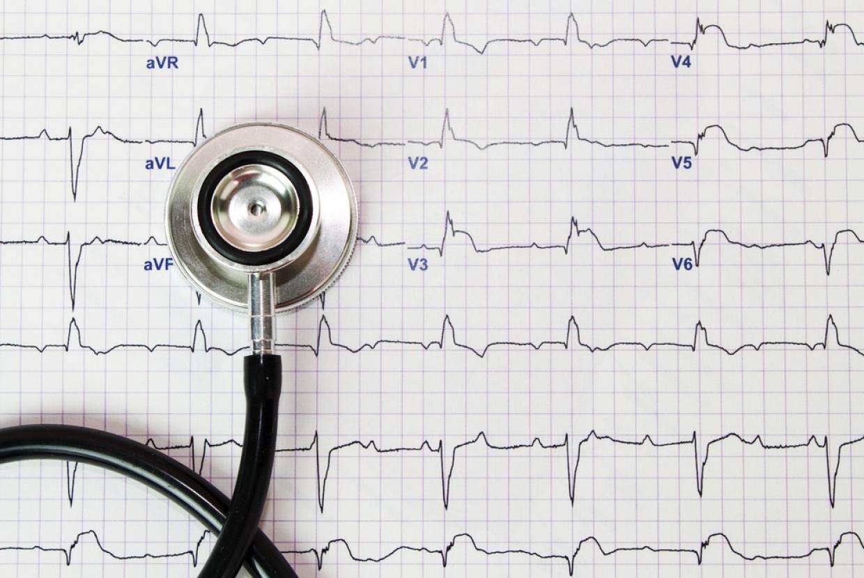
Electrocardiogram
An electrocardiogram (ECG) is a quick, painless test that measures the electrical activity and rhythm of your heart. The ECG will show a variety of lines and waves which will be analysed to see if there is reduced blood flow to the heart muscle or if you have had any recent damage to the heart. The ECG will also show how fast your heart is beating and any irregularities in its rhythm. Sometimes your doctor will compare your current ECG to previous ones. This helps to determine if changes to your electrocardiogram are recent. Since the ECG has limitations, your doctor may refer you on for further tests.
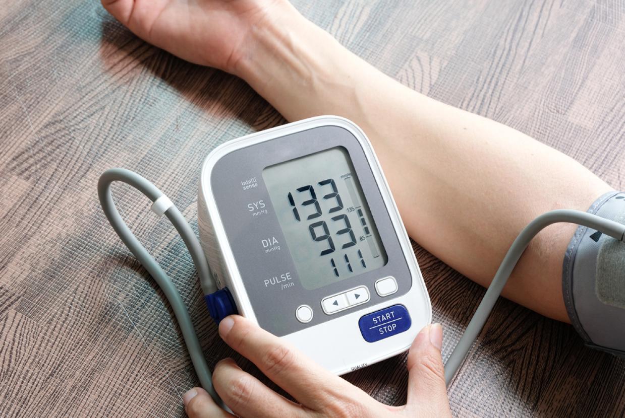
Exercise Stress Test
For many people who have significant narrowing of the arteries supplying the heart muscle, the electrocardiogram (ECG) recording made at rest can be normal. An Exercise Stress Test is an ECG recording made while the patient is exercising to see if their heart rate and rhythm is affected by activity or stress. During an exercise stress test you will walk on a treadmill while your ECG and blood pressure are monitored and recorded. The doctor will be looking at the ECG to see if any changes indicate coronary heart disease. The doctor will also be interested in how much exercise you are able to do and whether you experience chest pain or breathlessness.
Ambulatory Monitoring
Ambulatory monitoring involves monitoring your heart while you go about your normal daily activities. You will be asked to wear a small portable monitor at home.
A Holter monitor records the electrical activity of your heart while you do your usual activities. It is a small, portable ECG machine worn at home over a 24–48 hour period. Many symptoms like palpitations or fluttering in your chest become noticeable only during exercise, eating, stress, or even sleeping. A continuous recording is more likely to detect any abnormal heartbeats or rhythms that occur during these activities.
A Patient Activated Event Recorder is a device which records the ECG if you push the button when you feel your symptoms. This type of monitor may be more appropriate if your palpitations or fluttering sensations are less frequent.
Echocardiogram
An echocardiogram (echo) is a test in which ultrasound is used to examine the heart. The echo produces an image of the heart on a screen. It shows the structures of your heart including the 4 chambers and the valves. It assesses how well your heart is pumping, the presence of fluid around it, problems with your heart valves and information about the pressures within your heart.
A Transoesophageal Echocardiogram (TOE) is a special type of echocardiogram used when a closer and more defined image of the heart valves is needed. Pictures of your heart are taken by inserting a probe into your gullet (oesophagus). These pictures are clearer because the oesophagus is close to your heart and the chest wall is not in the way. The back of your throat will be sprayed with some numbing solution and although you will be awake during the procedure, you will be given some medication to make you feel relaxed and sleepy.
Cardiac Catheterisation
Cardiac catheterisation, also known as an Angiogram,is a procedure in which a small plastic tube is placed within a large artery in your groin and fed to the arteries in your heart. A special contrast agent (dye) will then be injected through the catheter and a series of x–rays will be taken. This technique is used to take pictures of the coronary arteries and the pumping action of the heart. It provides the most detailed and accurate information on the condition of your coronary arteries. Local anaesthetic is given in your groin or wrist before insertion of the catheter. You may feel pressure as the tube is inserted, as well as a warm sensation throughout your body when the dye is injected. The procedure generally lasts for less than 30 minutes. Afterwards you will be asked to lie still for 3–4 hours to allow the puncture site in the groin to heal (this is only if the groin is used).
Electrophysiological Studies (EPS)
If you have abnormal heart rhythms (arrhythmia) or palpitations you may be referred for this test. As with an angiogram, fine tubes (electrode catheters) are fed into a vein and/ or artery, usually in the groin. They are then gently moved into the heart, where they stimulate the heart and your heart’s electrical activity is recorded. This helps the doctors determine why you have the abnormal rhythm and decide on the best form of treatment.
Content supplied by...