Diagnosing Cervical Cancer
If you have symptoms of cervical cancer, you usually start by seeing your GP (local doctor). They will examine you and may refer you to the hospital for a specialist assessment and tests. If your GP thinks you may have cancer, they will refer you urgently to the hospital.
Cervical cancer may also be diagnosed during cervical screening or after an abnormal smear test. This is not common but sometimes happens.
Tests to diagnose cervical cancer
To diagnose cervical cancer, you usually have the following tests:
Colposcopy
This test uses a microscope called a colposcope to look closely at the cervix. It shows any abnormal areas of the cervix and how abnormal these are.
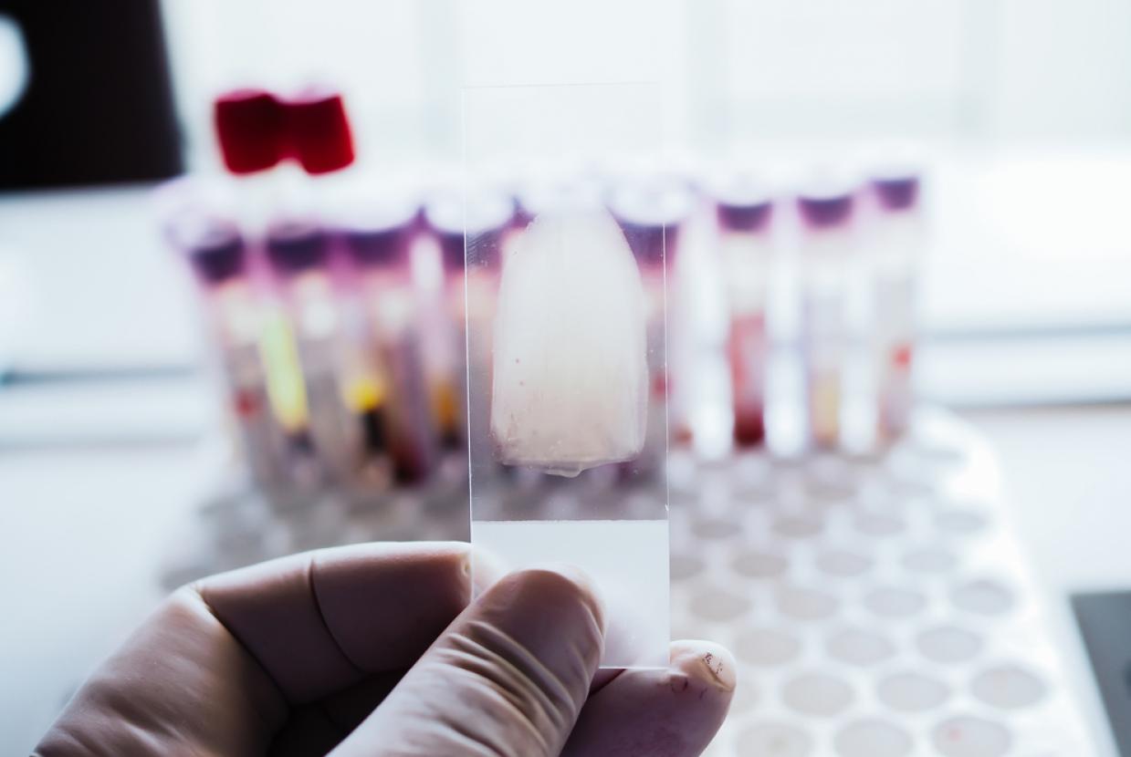
Biopsy
Samples of cells (biopsies) are taken from the cervix. You may have these taken during the colposcopy, or during another test such as LLETZ (Large loop excision of the transformation zone), NETZ (needle excision of the transformation zone) or cone biopsy. After the test, the samples are checked at a laboratory for cancer cells.
It is normal to have some bleeding or discharge after a biopsy. Your doctor or nurse will explain what to expect. They may advise you not to have penetrative sex, use tampons or go swimming for a time. This is to reduce the risk of infection and to give the cervix time to heal.
Biopsy results may take 2 or 3 weeks. Ask your doctor or nurse when you will get the results. Sometimes a LLETZ or cone biopsy removes all the cancer cells from the cervix. For very early-stage cervical cancer, this may be the only treatment needed.
Further tests
If the colposcopy or tissue collected during treatment for abnormal cells show you have cervical cancer, you will need to check:
- whether the cancer has spread beyond the cervix (called staging)
- your general health.
These tests help your doctors plan your treatment. You may have some of the following tests:
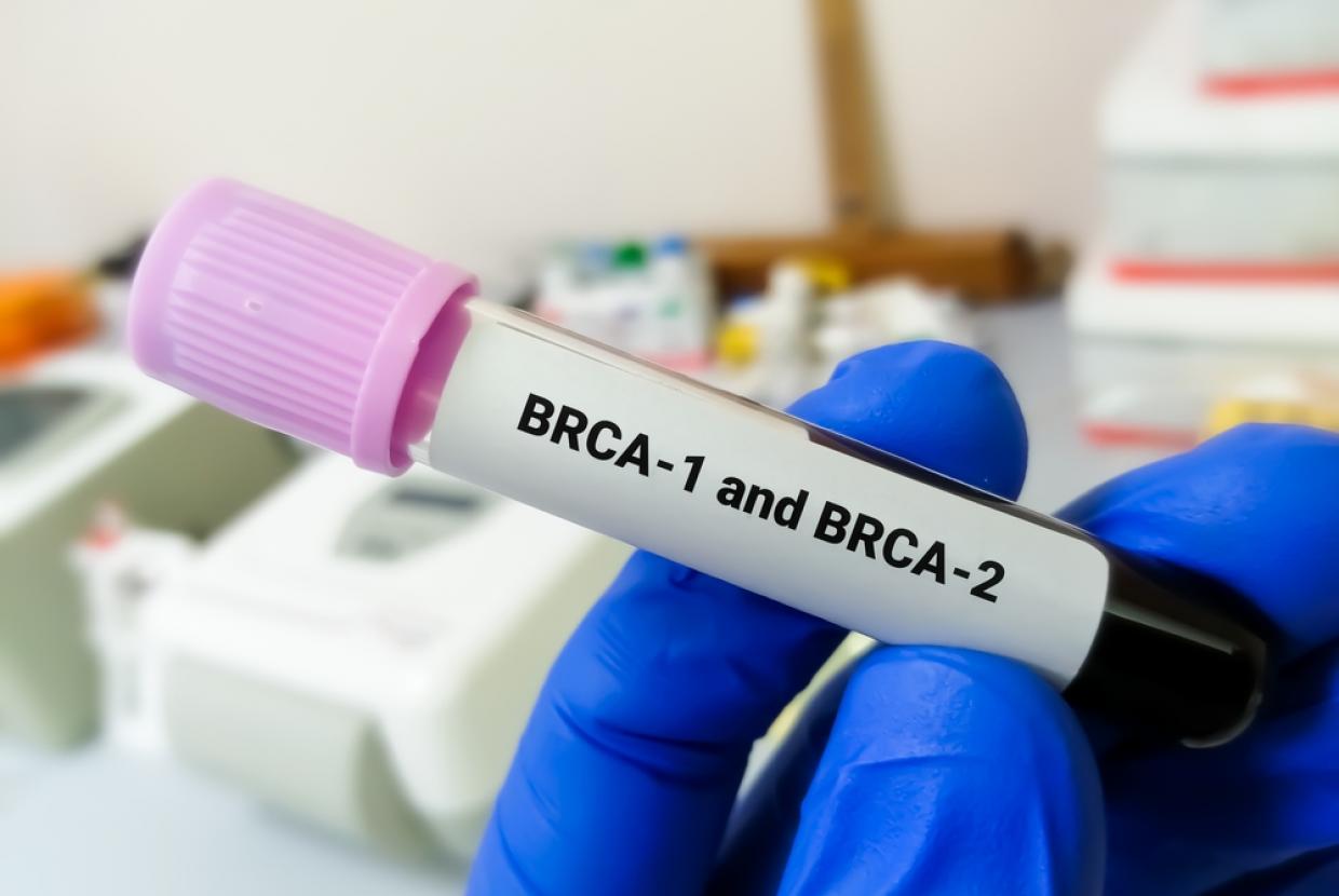
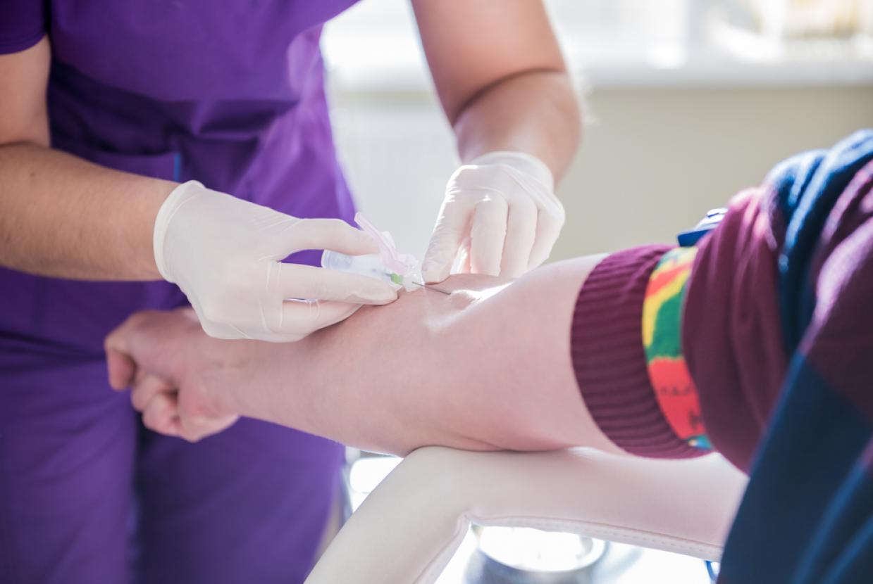
- Blood tests - Blood tests can check your general health and the number of blood cells in your blood (blood count). They can also check how well your kidneys and liver are working.
- Chest x-ray - This uses x-rays to take a picture of your chest. It may be done to check your general health and to look at your lungs and heart.
- MRI scan - An MRI scan uses magnetism to build up a detailed picture of the inside of your body.
- CT scan - A CT scan makes a three-dimensional (3D) picture of the inside of the body using x-rays taken by the CT scanner.
- PET or PET-CT scan - A PET scan uses low-dose radiation to check the activity of cells in different parts of the body. It is sometimes given together with a CT scan (PET-CT scan).
- Examination under anaesthetic (EUA) - Sometimes a test called an examination under anaesthetic (EUA) is needed, but this is not used very often. This is an examination of the vagina and cervix that is done under a general anaesthetic.
Content supplied by...